1. Prepared by Regina Kwon, MD, MPH
Reviewed by Patricia Simner, PhD
With thanks to Maritsa Angelou and Hillary Ajife for their technical assistance.
An elderly patient with a history of hypertension and hyperlipidemia presented to the Emergency Department (ED) with diaphoresis, lightheadedness, and pain in the epigastrium and lower chest. They had no trouble breathing. An electrocardiogram (ECG) was nonspecific. They were given Tylenol, nitroglycerin, and ondansetron, without improvement. The patient's white blood cell count was 19 K/cu mm (ref., 4.5-11), and a troponin I test, repeated three times, was negative. CT imaging of the abdomen and pelvis showed no significant abnormalities. Cardiac angiography revealed three-vessel disease.
The patient underwent percutaneous catheterization, with placement of a drug-eluting stent in the left anterior descending artery. They continued to have abdominal pain but felt better after several bowel movements. The white blood cell count remained elevated (max, 18.5 K/cu mmm), but the patient had no fevers. They were discharged after a one-week stay and given prescriptions for clopidogrel and low-dose aspirin.
Two weeks later, the patient returned to the ED, describing two days of stabbing right-sided chest pain. An ECG and a troponin I test were negative. They had mild leukocytosis and an elevated D-dimer level. CT imaging showed emphysematous cholecystitis. Due to the recent stent placement and risk of clotting, the patient was not a candidate for cholecystectomy. Instead, interventional radiology placed a percutaneous cholecystostomy tube and drained the fluid, which was sent to the Microbiology laboratory for aerobic and anaerobic bacterial cultures.
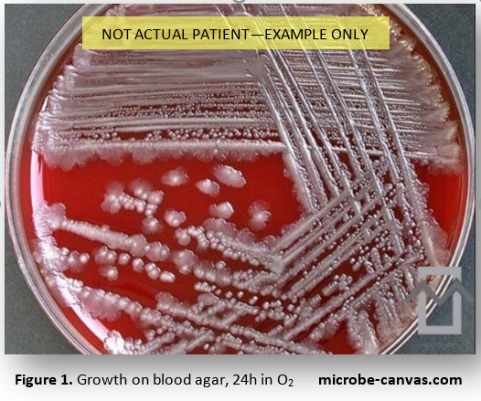
A Gram stain of the fluid identified very light polymorphonuclear leukocytes and very light gram-negative bacilli. The next day, heavy growth of two different morphologies was observed (Fig. 1) on the aerobic and anaerobic plates. Both morphologies were oxidase-negative and catalase-positive. The cultured samples were submitted for matrix-assisted laser desorption/ionization time-of-flight mass spectrometry (MALDI-TOF MS) analysis.
Which organism could this be?